CheckEmoji Community · the emoji forum
🏠 Home 🆕 What's new ❓ Unanswered 🔥 Popular 📡 RSS Members 👥 0 online log in · register
Home › Nicholas Myers › Posts

Posts by Nicholas Myers

1304 posts shown.

Brenda Parker5 said:The symptoms for RLS are exactly the same for me—honestly, I feel way better when I'm moving around. If that crawling sensation, tingling, and constant urge to move isn't RLS, then I must have been dreaming during my half-hour chat with a neurologist from the Mayo Clinic.

Based on what’s being said here, it’s clear you haven't actually been diagnosed with RLS. Yet, you're offering advice by framing it as if you're managing this and other conditions, which is just misleading everyone else. It's unacceptable.

I’m asking politely: please stick strictly to the topic you started.
Brenda Parker5 said:Alpha-lipoic acid is only stable in the R-ALA form—so if you weren't taking that, you basically threw your money down the drain. Btw, R-ALA combined with ALCAR is absolute magic.
My neurologist prescribed me Pregabalin 75mg for BFS (benign fasciculation syndrome) and potential SFN (small fiber neuropathy). You can't officially prove SFN unless you do a biopsy.

I deal with fasciculations, twitching, tingling, crawling sensations, all that good stuff...

Between the Pregabalin, R-ALA, ALCAR, and benfotiamine, I feel like a new person. Maybe do a little homework before jumping to conclusions.
With just Pregabalin alone for four months, maybe I felt 15% better at most. With this combo, though? It was a literal game-changer.
I also started a B-complex—specifically B12 and folate in their methylated forms.
advanced forms of B-vitamins, including benfotiamine (thiamin/vitamin B1), pyridoxal 5-phosphate (P5P/vitamin B6), methylcobalamin (vitamin B12), patented Pantesin(R) (pantethine) and 5-methyltetrahydrofolate (folate). These superior forms offer higher bioavailability for reliable supplementation.

This was a randomized, double-blind trial conducted in a hospital setting. A total of 64 consecutive patients (mean age 61 years; range 29-85) with acute backache and moderate sciatica were recruited. The 33 patients in group received 1180 mg of ALCAR daily for two months and the 31 patients in group 2 received 600 mg of R-ALA daily for the same period. The researchers measured changes in clinical signs and symptoms as measured on the Neuropathy Impairment Score in The Lower Limbs (NIS-LL) questionnaire, the Neuropathy Symptoms and Change in The Lower Limbs (NSC-LL) questionnaire, and the Total Symptom Score (TSS) questionnaire. The next level of improvement was how successful were the supplements in improving neurological deficit (as measured by electromyography) compared with baseline.

Both treatments showed significant improvements from the start regarding neuropathy (nerve pain) on electromyography by day 60. While the ALA group saw slightly better mean improvements than the ALCAR group, the difference between the two wasn't statistically significant. Notably, 71% of those on ALA said they needed fewer painkillers, compared to 45.5% of those on ALCAR (p < 0.05). The study is published in The Journal of Clinical Drug Investigation, 2008;28(8):495-500. Commentary by Jerry Hickey, R.Ph.; research indicates that pairing ALA with ALCAR might boost results significantly. Furthermore, a recent study in the Journal of Cellular and Molecular Medicine suggests that combining ALCAR with ALA enhances energy production, antioxidant activity, and cellular repair at concentrations 100-1000 times lower than when used individually in studies involving human brain cells and protection against Parkinson's disease.

Watch your tone. So far, from what I can see, everyone jumping into this thread is being completely off topic and hasn't offered a single valid argument. We aren't discussing back pain, Parkinson's, or neuropathy here!

As for suggesting or pushing supplements, I've already said: stick to the subject you actually opened this thread for.
I have no idea what you were trying to accomplish by quoting this study, but anyway...
The data shows that ALA yielded greater mean improvements compared to ALCAR. Even if the gap between the groups didn't hit that statistical significance mark...

That doesn't support the point you're trying to make. 🤷

And not a single word about RLS, which is the entire point of this thread! 🤔
steeleagle152 said:I’ve had blood work done before, but never this detailed. No one really sent me for it. My CRP has been consistently elevated since my splenectomy (following a car accident). It goes from 15 to higher, but usually hovers somewhere between 15 and 20. I don't smoke or drink. I am overweight, though I stay active through my job—it isn't a desk job. I only take medication for high blood pressure, and occasionally Praxiten (about three or four times a month) because I work three shifts; when I can't sleep after a night shift, I take one tablet. As for why the results look small... I don't know. Maybe it's just because I uploaded them via my phone to the forum.
Thanks for the clarifications.

Ma'am,

Elevated CRP levels shouldn't be linked to a splenectomy—that might happen in patients with thalassemia, but that doesn't apply here—rather, they indicate inflammation. Essentially, there is an inflammatory process occurring in the body. Granted, obesity can also trigger inflammation, which could reflect as an increased CRP level. Still, quite a bit of this is unusual. I suggest more comprehensive testing; the combination of elevated leukocytes, neutrophils, and CRP suggests an inflammatory cause that remains unclear based on what you've shared.
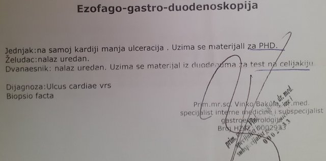
David Flores68 said:I think I was remembering things wrong regarding the whole mix-up with celiac disease and the samples. The doctor performing the endoscopy definitely took a duodenal biopsy (here is his report)...

image

...however, the only report I actually received from the pathologist is the one in the previous post, which is titled esophagus biopsy (even though it mentions the tissue corresponds to the stomach mucosa, but I assume the mucosa is similar in the esophagus) and there's no mention of the duodenum or celiac disease. Is it possible the pathologist processed the duodenal samples too, but I was mistakenly given only the report for the cardia tissue? I remember the technician searching for the results for quite a while; perhaps there were two papers (esophagus and duodenum), and she accidentally handed me just the esophagus one?

Now I see that I confused myself as well, and it’s actually a stomach biopsy rather than an esophagus one, despite what the title says.

Basically, the sample is stomach mucosa, so the changes aren't related to reflux—my apologies.

My answer regarding celiac disease still stands—the stomach or esophagus mucosa isn't where you'd see specific changes tied to celiac disease, so the pathologist wouldn't have commented on it. Since the gastroenterologist's report mentions a duodenal biopsy, you would expect the pathology report to describe the duodenal mucosa as well. If it's missing, it's possible that during the endoscopy, they didn't take a duodenal sample but instead focused on the stomach, since the pathology report refers to "stomach mucosa samples" in the plural. There is also the possibility that the findings were described separately, though it isn't standard practice to issue separate reports for samples taken during the same endoscopy.

Regarding the other part of your question: typically, to diagnose *H. pylori*, biopsies are taken from several different parts of the stomach, specifically the antrum and the corpus, because the bacteria can be unevenly distributed throughout the lining. Furthermore, your report describes intestinal metaplasia, and *H. pylori* is rarely found in areas with intestinal metaplasia. It prefers an acidic environment, whereas gastric acid secretion decreases in areas with intestinal metaplasia, making it less hospitable. So, it isn't unusual that only rare bacteria were noted in the report.

Intestinal metaplasia is a precancerous condition and increases the risk of developing stomach cancer. Regular follow-ups are necessary.

Diagnosing *H. pylori* via a stool antigen test is highly accurate. It is possible, though very rare, for that test to come back negative even if *H. pylori* is present in a biopsy. However, that is quite uncommon.

To clear up the dilemma of whether the bacteria in a biopsy is *H. pylori* or not, immunohistochemical analysis can sometimes help, but it is generally recommended to take a larger number of biopsy samples and look for *H. pylori* in samples where intestinal metaplasia is absent.

You should consult your gastroenterologist with these results to discuss monitoring and next steps.
steeleagle152 said:I’m not sick, nor have I been lately, but I’m wondering if these elevated values could be because I don't have a spleen? I had a splenectomy about 9 years ago.

Ma'am,

Given what you mentioned and following the guidelines in the opening post of this thread—where we ask for context regarding why tests were ordered, any existing conditions, medications, or even smoking habits—it’s clear that all these factors can shift lab results. Without that context, interpretation isn't accurate.

In short: there's no pathology in the liver panel. The GGT result is completely irrelevant here, and the cholesterol is only marginally off. Nothing to worry about yet, though suggesting lifestyle and dietary adjustments is always good practice.

Regarding the blood work, mild thrombocytosis is actually expected in someone who has undergone a splenectomy; such individuals may also show slightly higher leukocyte counts. You didn't mention the reason for the splenectomy (was it trauma?), and blood profiles can vary depending on why the organ was removed.

Typically, one sees mild leukocytosis (usually not exceeding 15 x 10^9/L), and the differential usually shows lymphocytosis (lymphocytes around 60%). In your case, however, neutrophilia is dominant. Neutrophilia can occur, and in most patients, it's transient and mild to moderate, though in some, it can persist longer. It's vital to know what your previous blood work looked like—have you had any tests in the last 9 years? We also need to consider your history: smoking, medications, physical activity levels, etc. All of this matters for an accurate reading.

And if I may ask, why is the photo so small? It's quite difficult to make out the numbers...
David Flores68 said:Look, two things are bothering me here:

1. why isn't there any mention of celiac disease?
I think I was sent for a gastroscopy because of suspected celiac disease, so why didn't the pathologist write anything about it?

Sir,

It’s not mentioned because celiac disease doesn't show up through specific changes in the histological findings of the esophagus—it shows up in the small intestine. Changes in the esophagus related to celiac aren't specific; they can look just like changes caused by acid reflux.

2. This last line regarding H.p.
"rare bacteria similar to H.p."

Why "similar" to H.p.? Does that mean the bacteria looked like H.p. but weren't actually H.p.? Or is this just standard phrasing in a pathology report?

Strictly speaking, there are other groups of spiral bacteria that look very much like Helicobacter pylori—take Helicobacter felis or Helicobacter heilmannii, for example. The pathologist is clearly being cautious, making it clear that the microorganisms they observed might not actually be Helicobacter pylori. In this context, "rare" means they were few in number. Therefore, based solely on this finding, it's impossible to say with absolute certainty whether we're dealing with an infection caused by Helicobacter pylori or something else.
jaderanger6 said:My gastroenterologist wrote this under the diagnosis on my results:

SIC
Gastropathy vs.

I figured out the "vs." part means Irritable Bowel Syndrome, but what exactly is gastropathy?

"Gastropathy" is just a broad medical term for stomach issues, and "vrs." is a Latin abbreviation that translates to probably.

jaderanger6 said:The doctor ordered immunoelectrophoresis for proteins along with several other tests. Looking at this mountain of paperwork, I can't tell if I actually got the immunoelectrophoresis done. Can anyone tell me if "(S) Monoclonal protein - immunotyping" is the same thing?

Monoclonal protein - immunotyping is an immunofixation test. Immunoelectrophoresis itself involves measuring IgA, IgM, and IgG. It’s likely the doctor ordered both, but they aren't the exact same procedure.
Since we haven't established a fever pattern, the illness has been dragging on for two weeks. Lab results are trending downward, and the antibiotic clearly isn't cutting it—we should have seen improvement within a 72-hour window. In my view, hospitalization is the only real path forward. I routinely admit patients with this exact profile. Even the initial urinalysis didn't point toward a urinary tract infection.
Frank Anderson9 said:Yeah, her primary care doctor. She went through the ER, but they didn't really find anything significant, sent her home, and just prescribed an antibiotic.

At a hospital ER?
Frank Anderson9 said:Hi everyone,
my wife's mother has been doing poorly for two weeks now.

To whom it may concern,

Was this procedure ordered based on a recommendation from her primary care physician?

Given these results and how long she's been suffering, hospitalization seems necessary.
Is NAC a miracle supplement? in Health ·
Brenda Alvarez24 said:Where did you pull that quote from that you sent me in a DM? Honestly, feel free to post that in the SLE thread so everyone dealing with lupus can see it—it shouldn't be a secret, right?

No. I am asking politely that discussions regarding this supplement stay EXCLUSIVELY within this thread, rather than bleeding into every single discussion on the Health board!

Once and if this supplement actually makes it into the official treatment guidelines for lupus, MS, or other conditions, then we can talk about it in those threads. Until then—unless we are discussing managing a productive cough or acetaminophen toxicity—keep the conversation about this specific supplement right here.

Thanks.
David Flores68 said:Could someone please help me interpret these results?

image

I'm 33. My doctor ordered this panel because of some nodules popping up on my right fingers, though I've been dealing with undiagnosed GI issues for years now. Honestly, I can't tell if she's looking into the first or the second.

As far as I can tell, these other results fall within the normal range. Still, they might be saying something significant—perhaps ruling certain things out. Here they are:
imageI can't see any images yet. Go ahead and upload the photo whenever you're ready.

To whom it may concern,

Normal calprotectin levels, paired with steady CRP and ESR readings, suggest that your GI issues likely aren't stemming from inflammatory bowel disease. That’s the takeaway from this specific set of data. However, it doesn't rule out other culprits; it just means we can't point to IBD as the cause based on these numbers alone.

Low vitamin D levels signal a deficiency that needs addressing with supplementation. Given how low these numbers can get, the most effective approach is typically a high-dose regimen—something like 50,000 units once a week for about six to eight weeks—before transitioning to a standard maintenance dose. In the US, you'd likely see a prescription for an oral solution like D-vital to get things back on track.

Regarding those nodules, this specific result doesn't point to a clear culprit just yet. One possibility could be calcium depletion stemming from a long-term Vitamin D deficiency—which might trigger secondary hyperparathyroidism—though that doesn't strike me as particularly likely in your situation. Still, once we review everything, it would be worth considering whether checking your calcium, PTH, and magnesium levels makes sense. Ultimately, a physical exam and a biopsy remain the gold standard for getting a definitive diagnosis.
Carl Thompson26 said:Hello,

I’d appreciate some insight. 36 years old, healthy, fully vaccinated (two doses), never had COVID. Had blood work done in January; results were:

WBC 2.7
NEUTROPHILS 1.60
MONOCYTES 0.30

Results repeated in February and March. They remain consistent.

April 14, 2022
DKS CYTOLOGY REPORT:

eosinophil granulocytes 1%
segmented neutrophil granulocytes 54%
lymphocytes 31%
reactive lymphocytes 10%
monocytes 4%

ER: mild anisopoikilocytosis, normochromia
GR: normal morphology
TR: present, normal morphology

Recommendation: hematological follow-up.

Hello,

From what I can see, you have lymphopenia, which warrants further investigation.

The first question is how your blood counts usually look—I assume you've had labs done at some point in your life. In other words, is this low white cell count a new development from January 2022, or have your levels always sat at the bottom end of the normal range? Have you been sick at all in the last six months? Is it possible you had an infection, a fever, or just felt unwell recently? Have you had any medical exams lately? Any high-risk sexual encounters? Are you taking any medications or consuming alcohol? What about your weight—has it shifted significantly in the last six months? Have you dealt with any serious infections? Fevers, night sweats? Rashes or skin changes?
brightscout9 said:To whom it may concern,

Ponovi sam nalaze koje sam radio prije cca 4 mjeseci

I'd appreciate your thoughts on this...

Thanks.

https://amaranth-dorris-41.tiiny.site/

To whom it may concern,

Transferin saturation remains high at 75%, even with the recent spike in creatine kinase. Given these results, it’s time to touch base with the primary care physician to figure out the next steps for testing.
Quote : EL Gato Félix
Emily Ortiz27 said:Thank you. Much appreciated.
Instead of waiting for an actual follow-up appointment with the immunologist, I stumbled upon the girl's lab results on a health portal.

image

This report was issued without any follow-up communication or a proper clinical review of my child. On top of that, the findings completely contradict the guidelines you provided. How do you suggest I proceed from here?
Should I just stick with the results, or reach out for a second opinion at Mayo Clinic?
I currently have an active referral for the post-COVID clinic in Miami, so I’m not entirely sure if I could even use a different referral to see the same specialist at another facility.
And who exactly should I contact over in Miami?
The little girl isn't running a fever every single day anymore. It fluctuates—sometimes she’s back to normal, sometimes it hits 98.6°F, and other times it creeps up a bit higher.
Abdominal pain persists. It’s more pronounced following physical activity.
How should we approach physical activity here? He’s training three times a week, and that doesn't even account for his regular PE classes at school.

Dear Madam,

I wrote my initial assessment based on the specific history provided: a young girl referred to a post-COVID clinic following prolonged fever, where she underwent an immunology consultation. My response was simply outlining the necessary diagnostic steps for anyone presenting with persistent fever and the specific lab results you shared.

Now that the fever has broken, my answer to that question would look quite different.

One fact remains, though: the reasoning behind ordering an ANA test. If the girl didn't have a fever at the time she saw the immunologist, the motivation for that specific lab work is unclear. It’s a well-documented phenomenon that ANA titers can spike—sometimes even reaching high levels—months after a SARS-CoV-2 infection or other viral bouts, such as mononucleosis. But you don't just run an ANA for those conditions. You order it when there is actual clinical suspicion of systemic connective tissue diseases, like lupus, lupus-related disorders, or certain autoimmune liver issues. In almost any other scenario, the test provides zero value for diagnosis or prognosis. To be blunt, it's an unnecessary expense. I fail to see why the immunologist felt this test was warranted.

Now that the test results are in—results that show a deviation—I have to wonder: which clinician looked at the clinical picture, factored in the history of a prior SARS-CoV-2 infection, and arrived at a conclusion that actually made sense? I’m circling back to my original question: what role did the ANA play in this entire diagnostic process? Why was it ordered in the first place, and how exactly did it guide the physician toward their final conclusion? I’d be interested to hear some actual reasoning on that.

Second, I have to disagree with how they handled the data—or rather, the lack of action regarding it. Specifically, the note that "abdominal pain persists and intensifies with movement." So, we have a child reporting stomach pains, yet no abdominal ultrasound was even requested? Honestly, I can't wrap my head around why.

Since things seem to be settling down and resolving on their own, most of those tests I mentioned earlier probably aren't strictly necessary. That said, if the abdominal pain persists, I’d suggest getting an abdominal ultrasound just to be safe.

Blood cultures probably aren't worth the effort at this stage. However, if that abdominal pain persists, I’d still suggest running a serum protein electrophoresis, checking the albumin/creatinine ratio from a urine sample, testing stool for occult blood three times, getting a calprotectin test, and scheduling an abdominal ultrasound. Ultimately, though, my decision would hinge on the physical exam and the clinical picture.
Emily Ortiz27 said:Thank you so much!
What you're recommending seems quite extensive.
We're still waiting for an opening to even see an immunologist, so we'll have to see what's possible. Given how broad this testing panel is, do you think there are specific areas we should focus on or push for if needed?
Unfortunately, our experience here is that these kinds of situations are handled much more modestly.
If things don't work out, could you suggest a place where we might get this level of diagnostic work done?
It doesn't matter if it's in Washington, D.C.; almost all of the doctors for my other child are based in Washington, D.C. anyway.

Ma'am,

if the child is still running a fever, I believe all those tests are necessary. It's a different story if the fever has subsided.

If you can't get these tests done locally, regarding a child with these specific issues, you can make an appointment at the pediatric rheumatology clinic at Children's Hospital.
Rachel Davis said:They sent me in for a urine culture because my white blood cell count was up and they found bacteria in my urine, but I can't make sense of this report:
leukocytes positive (~15), erythrocytes and nitrites negative. Microorganism count 10^4 CFU/ml. Resident flora of the distal urethra.

There are no signs of a urinary tract infection.
Emily Ortiz27 said:I've lost track of what else might be missing from the list. I attached everything that's been completed to my previous post.
When we call them up, the response is always the same: everything is still being processed, so there's no way to schedule any follow-up consultations just yet.
No abdominal ultrasound was performed, nor any of the other tests you mentioned.

Dear Madam,

Here’s what I’d suggest adding to the list: anti-dsDNA, anti-SS-A, CH50, LAC, beta-2-GPI, and both IgM and IgG anticardiolipin antibodies. You should also look into ANCA, serum protein electrophoresis, and a urine albumin/creatinine ratio. For basic labs, check CK, PV, APTV, fibrinogen, D-dimer, bilirubin, and an electrolyte panel including Na, K, Cl, Ca, Mg, and P. Don't forget a peripheral blood smear, three stool tests for occult blood, and calprotectin. On the imaging side, get an abdominal ultrasound, a chest X-ray, and an echocardiogram. Once those results are in, we can decide if further testing is needed based on how the symptoms present. Finally, it might be worth running serology for EBV, CMV, Bartonella, and a QuantiFERON test.

I'll wait until all the results are in—bloodwork, endocrinology, OBGYN—then we can actually see what the full picture looks like.
If the cause is idiopathic, what does the treatment plan actually look like?

Treatment follows a similar logic to managing hyperprolactinemia—think along the lines of taking Bromergon 1,25 mg to 2.5 mg daily, or perhaps Dostinex 0,25 mg once or twice a week.
Paul Ramirez59 said:I’ve stopped taking my PPI after talking things over with my gastroenterologist, though he’s skeptical about the link to the medication. He’s leaning toward Irritable Bowel Syndrome instead. The thing is, I don't get this vomiting with IBS—we both know that—but he isn't budging. When I brought up a stool cytology test, he just brushed it off, saying I'd already done it. From what I recall, I had tests for bacteria, viruses, and parasites. Can anyone explain what a stool cytology actually looks for? What are they looking at under the lens?

It basically checks for the presence of inflammatory cells under the microscope.

