I feel like we’re seeing these massive, club-record fees more and more lately, even for players who aren't household names yet. It feels like such a huge gamble to drop that kind of cash on a single prospect. Do you think these big investments actually pay off long-term, or is it just chasing hype?
Home ›
David Flores68 › Posts
Posts by David Flores68
52 posts shown.
How to congratulate someone on a new baby?
in Miscellaneous ·
I’m a bit socially awkward—I often struggle to wrap my head around these kinds of unspoken social norms—so I figured I’d just ask: is it actually customary to offer congratulations when someone has a baby?
My cousin Smith just had a new baby; would this be an appropriate moment to send over a quick congratulatory message via WhatsApp?
My cousin Smith just had a new baby; would this be an appropriate moment to send over a quick congratulatory message via WhatsApp?
One tooth is way smaller than the others—what's the cause?
in Dentistry & Oral Hygiene ·
coppergardener said:I’m no expert, obviously, but I would definitely head to the dentist for a professional cleaning and some actual advice—those little pits on your molars look suspiciously like erosion to me.
Are you suggesting we're looking at erosion caused by stomach acid reflux?
One tooth is way smaller than the others—what's the cause?
in Dentistry & Oral Hygiene ·
I have one tooth that is roughly half—maybe even three-quarters—smaller than all my other teeth. It’s been this way for several years now; there was no injury involved, nor has there ever been any pain. I simply looked in the mirror one day and noticed just how much smaller it appeared compared to its neighbor. As for the shape of the biting surface, it looks perfectly normal to me—nothing suggests significant wear or erosion.
Does anyone happen to know how this could have happened or what the underlying cause might be?
I've attached some photos below. I wrote the description in English because I actually posted this same question on an American forum recently, though I didn't receive a single helpful response. My apologies if the photos are subpar; it's surprisingly difficult to get a clear shot that stays in focus while also capturing the right angle to demonstrate this size discrepancy.
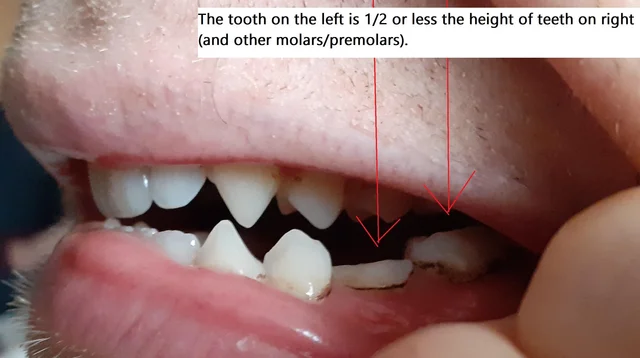
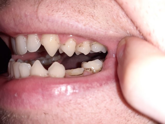
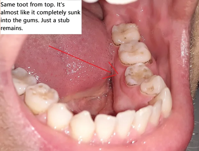
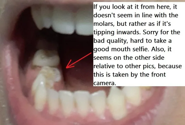
Does anyone happen to know how this could have happened or what the underlying cause might be?
I've attached some photos below. I wrote the description in English because I actually posted this same question on an American forum recently, though I didn't receive a single helpful response. My apologies if the photos are subpar; it's surprisingly difficult to get a clear shot that stays in focus while also capturing the right angle to demonstrate this size discrepancy.
Maria Fisher46 said:Dear Sir/Madam,
This question really ought to be directed toward the pathologist who actually examined the specimen. I can't speak to what they saw firsthand, though I can certainly infer from the wording of the report that they identified gastric mucosa. Essentially, the pathologist believes the sample consists of stomach lining rather than esophageal tissue. That presence of stratified squamous epithelium on part of the surface? That could simply be due to the "Z-line"—that specific junction where the distal esophagus meets the cardia of the proximal stomach. It’s entirely possible that some esophageal mucosa was caught up in the biopsy during the procedure.
That said, the definitive explanation needs to come from the pathologist who reviewed and documented the findings. I simply cannot tell you what they observed—or what they *should* have observed.
Thanks!
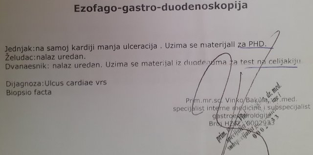
I’m feeling a bit thrown by my biopsy results. I recently underwent a gastroscopy, and the findings were as follows:
And here is the report from the pathologist...
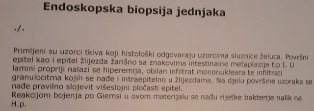
What’s really tripping me up is this—the pathologist's report states the material is consistent with gastric mucosa, yet it also mentions that part of the sample contains normal stratified squamous epithelium. From what I’ve gathered during my own research, the stomach lining should be composed of simple columnar epithelium, right?
Does this mean we're actually looking at an esophageal sample, and if so, could this intestinal metaplasia indicate Barrett's esophagus?
And here is the report from the pathologist...
What’s really tripping me up is this—the pathologist's report states the material is consistent with gastric mucosa, yet it also mentions that part of the sample contains normal stratified squamous epithelium. From what I’ve gathered during my own research, the stomach lining should be composed of simple columnar epithelium, right?
Does this mean we're actually looking at an esophageal sample, and if so, could this intestinal metaplasia indicate Barrett's esophagus?
Routine checkup vs. suspected cavity: what should I do?
in Dentistry & Oral Hygiene ·
I’ve noticed this dark spot on my tooth—I have a nagging suspicion it might be a cavity—so I went ahead and booked an appointment with my dentist. However, I only requested a routine checkup and didn't mention my specific concern about potential decay, which resulted in me being scheduled for the end of the month. Would I have secured an earlier slot if I had been upfront about having a specific issue rather than just asking for a standard cleaning? Is it possible to request an expedited appointment by citing the suspected cavity?
Maria Fisher46 said:Dear patient,
The samples collected during the second endoscopy weren't quite "high quality"—to put it bluntly, there simply wasn't enough material to draw any definitive conclusions based on the analysis provided. It’s also unclear why an IgA test was ordered back in November 2014, given that you have an isolated IgA deficiency, which makes those specific results irrelevant. I wouldn't go so far as to say celiac disease is ruled out—since the small intestine biopsy was inadequate for a proper analysis, and both the IgA and tTg IgA tests are essentially useless in your specific situation—but we can't be certain. Furthermore, the stomach biopsy was insufficient; it is entirely possible that areas of intestinal metaplasia still exist but were missed by the biopsy. Keep in mind, intestinal metaplasia isn't always visible to the naked eye during the procedure. As I believe I've mentioned previously, having normal calprotectin levels—especially alongside a normal CRP and ESR—suggests there is a very low probability that your gastrointestinal issues are linked to inflammatory bowel disease.
To be perfectly honest, looking at the findings presented, it was difficult for me to reach any meaningful conclusion.
Thank you so much!
Maria Fisher46 said:It looks like I've managed to confuse myself as well—it turns out we’re actually looking at a stomach biopsy, not an esophageal one like the title of the report suggests.
Actually, I need to correct myself—the sample was taken from the gastric mucosa, which means these changes aren't actually related to reflux. My apologies.
My previous response regarding celiac disease still stands—the stomach or esophagus lining isn't actually where you'd see those specific changes associated with celiac, which is why the pathologist didn't weigh in on that particular issue. Since the gastroenterologist’s report specifically mentions a duodenal biopsy, one would naturally expect the histopathology report to describe the duodenal mucosa as well. If that description is missing, there is a real possibility that during the endoscopy, they didn't actually take a duodenal sample at all, but rather focused on the stomach—especially since the pathologist’s report refers to "stomach mucosa samples," implying plurality. There is also the slight chance that the findings were documented in a separate report, though it’s certainly not standard practice to split up samples taken during a single endoscopy into different results.
Regarding the second part of your question—standard practice for diagnosing *Helicobacter* usually involves taking biopsies from several different areas of the stomach, specifically the antrum and the corpus, because these bacteria don't always distribute themselves evenly across the gastric mucosa. Furthermore, your results indicate intestinal metaplasia, and frankly, *Helicobacter* is rarely found in areas where intestinal metaplasia is present. To put it simply, the bacteria thrive in an acidic environment; however, in areas affected by intestinal metaplasia, gastric acid secretion decreases—which just isn't a hospitable environment for them. Therefore, it isn't at all surprising that the findings only show rare traces of the bacteria.
Intestinal metaplasia is essentially a precancerous condition—it bumps up the risk for developing stomach cancer. Because of that, you really can't afford to skip regular follow-up screenings.
Stool antigen testing for *H. pylori* diagnosis is highly accurate. It is possible—though extremely rare—to see a negative result on that test only to have *H. pylori* show up later in a biopsy. Still, that kind of discrepancy is quite uncommon.
When you're trying to settle the debate over whether a biopsy actually shows *H. pylori* or if it’s just a false lead, sometimes an extra step—like immunohistochemical analysis—can clear things up. That said, my advice is always to be thorough: grab a larger number of biopsy samples if you can, and specifically look for *H. pylori* in those areas where you aren't seeing any intestinal metaplasia.
With these results in hand, you really ought to schedule an appointment with your gastroenterologist—it’s best to sit down with them to coordinate your follow-up care and map out the next steps in your treatment plan.
Thanks for sharing your thoughts!
I have some additional lab results—I didn't want to dump them all here immediately because the post would have been massive, but I figured I'd share them now just in case they change the overall picture. There’s quite a lot to go through, so I’ll try to summarize the main points in bullet points and skip over the minor details:
After that gastroscopy—this was back on February 13, 2014—here is what came back.


February 20, 2014 — Negative stool test for Helicobacter.
February 28, 2014 — underwent a new gastroscopy.


So, here’s the deal—the HP didn't show up on this one. But, just like you pointed out earlier, they really ought to be taking biopsies from several different sections of the stomach, whereas here, they only grabbed a sample from one spot. On top of that, if I'm reading this correctly, the initial gastroscopy actually picked up HP at the cardia—yet, in this specific instance, they didn't even take a biopsy from the cardia.
November 18, 2014 — Ferritin and IgA levels both came back normal.
- Dec 18, 2014 - H. pylori came back negative again.
- March 11, 2016 - fT3, TSH, and fT4 were all normal.
- April 28, 2022.
Vitamin D was low (likely because I stay out of the sun far too much), and IgA and beta globulins were questionable.
ESR, CRP, protein levels, and calprotectin were all within normal range.
- I've had quite a few blood tests over the years, so I'll list them here.
- Symptoms:
There have been plenty of them over the years—constant gas every minute after eating, bloating and popping sounds in the gut (almost like tiny bubbles forming and bursting), stomach burning, a sensation of being overly distended if I eat too much followed by discomfort/pain, sensitivity to acidic foods (like apples, for instance), getting full way too quickly, and various other issues.
The symptoms have actually eased up over the years—they're very rare now—but the weight loss persists. My target weight is 65 kg (which I’ve maintained comfortably in the past, never dropping lower), yet since the onset of these symptoms, I haven't been above 60 kg. To me, this suggests that whatever the underlying issue was, it's still hanging around somewhere.
Regarding monitoring the metaplasia: since it's located—I assume—right where that ulceration used to be at the cardia, is it necessary to take another biopsy from the cardia to see if the metaplasia has progressed? Or is that something a doctor can just spot visually during a gastroscopy without needing a biopsy?
I remember asking about this on this thread once before, and someone mentioned that I could pull my lab results directly through the government portal—is that actually right?
Also, can I find older records there, say from back in 2013?
Also, can I find older records there, say from back in 2013?
Maria Fisher46 said:Dear sir,
Nothing is specified because celiac disease doesn't manifest through specific histological changes in the esophagus, but rather in the small intestine. Esophageal changes related to celiac disease aren't unique—they can easily mimic changes caused by acid reflux.
Strictly speaking, there are other groups of spiral bacteria that look remarkably similar to Helicobacter pylori—for instance, Helicobacter felis or Helicobacter heilmannii—and the pathologist was clearly trying to be cautious by noting that the microorganisms observed might not actually represent Helicobacter pylori. In this context, "rare" simply means they were few in number. Therefore, based solely on this finding, it’s impossible to say with absolute certainty whether we are dealing with an infection caused by Helicobacter pylori or something else.
Thank you!
Given that those bacteria were found within the ulceration at the cardia, is it possible to assume the bacteria caused the ulcer, rather than celiac disease or reflux?
What do you think the next steps should be, considering these results? I suspect those three stool tests for H. pylori came back negative precisely because we're looking at a different type of spiral bacteria (or perhaps the test is sensitive to all of them?). Isn't it essential to identify exactly which bacteria we're dealing with and eradicate them? My doctor hasn't prescribed any antibiotics, nor have they pursued the bacterial question any further.
Something occurred to me regarding this whole mess with the celiac findings and the samples. The physician who performed the endoscopy definitely took a duodenal biopsy (here is his report)...
...however, the only report I actually received from the pathologist is the one from my previous post, titled "esophagus biopsy" (even though it mentions the tissue corresponds to the gastric mucosa, I assume the mucosa is essentially the same in the esophagus). There is absolutely no mention of the duodenum or celiac disease. Is it possible that the pathologist processed the duodenal samples, but I was mistakenly handed only the report for the tissue from the cardia? I remember the technician spent quite a while searching for the results—perhaps there were two separate papers (esophagus and duodenum)—and she accidentally gave me just the esophagus one?
Something about my old lab results is really throwing me for a loop...
Ever since the summer of 2011, I’ve been dealing with these vague digestive issues. (The symptoms have fluctuated quite a bit over the years—diarrhea for a year straight, weight loss, and so on...)
On December 29, 2011, I did a stool test for Helicobacter, which came back negative.
Then, on November 7, 2013, I underwent a gastroscopy. It showed "minor ulceration" located right at the cardia. They took a biopsy of that ulceration, along with a sample from the duodenum because they suspected celiac disease.
The pathologist wrote this in the report:
Now, two things are specifically bothering me here:
1. Why isn't there any mention of celiac disease?
I'm pretty sure I was sent for the gastroscopy due to suspicions of celiac, so why didn't the pathologist note anything regarding that?
2. This last line regarding H.p...
"rare bacteria resembling H.p."
Why "resembling" H.p.? Does that mean the bacteria looked like Helicobacter but weren't actually it? Or is that just some standard medical jargon used in pathology reports?
If it actually *was* H.p., how is it possible that they found it now, when I had a negative result for H.p. two years prior (about six months after my symptoms first started)?
I realize those stool tests are roughly 90% accurate, so there's always a slim chance of a false negative. However, I went ahead and did that same stool test twice more after that (once on February 20, 2014, and again on December 18, 2014), and both times were negative. To top it off, I wasn't even prescribed any antibiotics for these bacteria following the biopsy.
So, if three different tests said there was no H.p., is it possible that the bacteria found during the biopsy were actually something else entirely?
And this part about "rare bacteria"—does "rare" mean there were very few of them present, or that they belong to a rare species of bacteria?
Ever since the summer of 2011, I’ve been dealing with these vague digestive issues. (The symptoms have fluctuated quite a bit over the years—diarrhea for a year straight, weight loss, and so on...)
On December 29, 2011, I did a stool test for Helicobacter, which came back negative.
Then, on November 7, 2013, I underwent a gastroscopy. It showed "minor ulceration" located right at the cardia. They took a biopsy of that ulceration, along with a sample from the duodenum because they suspected celiac disease.
The pathologist wrote this in the report:
Now, two things are specifically bothering me here:
1. Why isn't there any mention of celiac disease?
I'm pretty sure I was sent for the gastroscopy due to suspicions of celiac, so why didn't the pathologist note anything regarding that?
2. This last line regarding H.p...
"rare bacteria resembling H.p."
Why "resembling" H.p.? Does that mean the bacteria looked like Helicobacter but weren't actually it? Or is that just some standard medical jargon used in pathology reports?
If it actually *was* H.p., how is it possible that they found it now, when I had a negative result for H.p. two years prior (about six months after my symptoms first started)?
I realize those stool tests are roughly 90% accurate, so there's always a slim chance of a false negative. However, I went ahead and did that same stool test twice more after that (once on February 20, 2014, and again on December 18, 2014), and both times were negative. To top it off, I wasn't even prescribed any antibiotics for these bacteria following the biopsy.
So, if three different tests said there was no H.p., is it possible that the bacteria found during the biopsy were actually something else entirely?
And this part about "rare bacteria"—does "rare" mean there were very few of them present, or that they belong to a rare species of bacteria?
Maria Fisher46 said:Dear patient,
The normal calprotectin levels, along with a normal CRP and ESR, suggest there is a low probability that your gastrointestinal issues are linked to inflammatory bowel disease. That is what can be inferred from that specific portion of the results. However, we cannot determine the actual cause of these symptoms based solely on this data.
Your low vitamin D concentrations indicate a deficiency that requires treatment with vitamin D supplements. Given how extremely low your levels are, it would be best to start treatment with 50,000 units weekly for about 6 to 8 weeks, then transition to a maintenance dose. For a prescription, an oral solution like D-vital would work.
Regarding the nodules, this finding doesn't provide any clues as to what might be causing them at this stage. One possibility could be calcium deposition resulting from prolonged vitamin D deficiency—which could trigger secondary hyperparathyroidism—though I don't suspect that is likely in your case. Still, after a physical exam, it might make sense to check your calcium, PTH, and magnesium levels. Ultimately, a clinical examination and a biopsy of the lesions remain the gold standard diagnostic methods.
Many thanks!
Jacob Lopez51 said:You’re dealing with chronically low Vitamin D—which brings its own baggage—and one of the biggest fallout effects is an automatic drop in glutathione levels.
Just start supplementing with 5,000 units of D3 paired with K2.
Does this low Vitamin D level point toward anything regarding my gastrointestinal issues?
Can I just fix this by getting some sun instead of relying on supplements?
Could someone please help me interpret these results?

I’m a 33-year-old male. These tests were ordered because I've been developing these nodules on the fingers of my right hand—but I also have a history of undiagnosed gastrointestinal issues that have been hanging over me for years. I'm honestly not sure if my doctor requested this panel because of the skin symptoms or the gut problems.
As for the rest of the findings, they seem to fall within the standard reference ranges, though I wonder if they might still be hinting at something—perhaps ruling certain things out. Here they are:
 upload photo
I’m a 33-year-old male. These tests were ordered because I've been developing these nodules on the fingers of my right hand—but I also have a history of undiagnosed gastrointestinal issues that have been hanging over me for years. I'm honestly not sure if my doctor requested this panel because of the skin symptoms or the gut problems.
As for the rest of the findings, they seem to fall within the standard reference ranges, though I wonder if they might still be hinting at something—perhaps ruling certain things out. Here they are:
upload photo
Keyboard recommendations?
in Computer Peripherals ·
Does anyone have a recommendation for a budget-friendly wired keyboard that features play/pause controls positioned on the left side—somewhere easily reachable by my left hand?
How to do a home stool sample test
in Health ·
I’ve been browsing some videos on YouTube regarding how to collect a stool sample at home, and I came across this one American video showing a specific collection kit. It includes this little device—basically a mesh or a catcher—that you place over the toilet bowl to catch the specimen so it doesn't fall directly into the water. Does anyone know if there is anything similar available for purchase here in the States?
The sample I need to provide is specifically for a calprotectin test. Does it actually matter if the stool hits the water or the toilet bowl itself? I'm wondering if that would somehow contaminate the sample and make it useless for this particular screening, or is it perfectly fine to collect it even if it ends up touching the water?
The sample I need to provide is specifically for a calprotectin test. Does it actually matter if the stool hits the water or the toilet bowl itself? I'm wondering if that would somehow contaminate the sample and make it useless for this particular screening, or is it perfectly fine to collect it even if it ends up touching the water?
Has anyone actually managed to get through to the central ordering desk at Mayo Clinic over the phone?
I've tried calling both numbers listed here multiple times now, but it’s just endless ringing and nothing else.
I've tried calling both numbers listed here multiple times now, but it’s just endless ringing and nothing else.
I went in for blood work at my local community clinic back in February of last year, and they were using a system where the results were sent directly to my doctor—so I don't actually have a physical copy of those labs here. Is there someone I can email or call to request that they send a copy of those old results to my personal email address?
I was there about two or three weeks ago, and it wasn't crowded at all. There were maybe four or five people sitting near me, and I was in and out in about 15 to 20 minutes.
coppergardener said:Has anyone been to see a neurologist over at the Mayo Clinic recently? I’m trying to get a sense of how long the wait times are and what the crowds are like in the waiting room. (I'm actually not sure which specific doctor I'd be seeing yet.) Thanks in advance!
I was there about two or three weeks ago, and it wasn't crowded at all. There were maybe four or five people sitting near me, and I was in and out in about 15 to 20 minutes.
Maria Fisher46 said:No, there are still quite a few other tests and diagnostic steps required to rule out rheumatic disease—so my answer is that, at this stage, we cannot say that a rheumatic condition isn't part of the underlying cause of your symptoms.
Much appreciated!