#3561 ·
David Flores68 said:I think I was remembering things wrong regarding the whole mix-up with celiac disease and the samples. The doctor performing the endoscopy definitely took a duodenal biopsy (here is his report)...
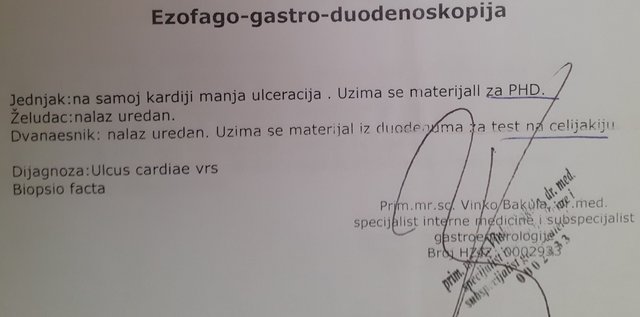
...however, the only report I actually received from the pathologist is the one in the previous post, which is titled esophagus biopsy (even though it mentions the tissue corresponds to the stomach mucosa, but I assume the mucosa is similar in the esophagus) and there's no mention of the duodenum or celiac disease. Is it possible the pathologist processed the duodenal samples too, but I was mistakenly given only the report for the cardia tissue? I remember the technician searching for the results for quite a while; perhaps there were two papers (esophagus and duodenum), and she accidentally handed me just the esophagus one?
Now I see that I confused myself as well, and it’s actually a stomach biopsy rather than an esophagus one, despite what the title says.
Basically, the sample is stomach mucosa, so the changes aren't related to reflux—my apologies.
My answer regarding celiac disease still stands—the stomach or esophagus mucosa isn't where you'd see specific changes tied to celiac disease, so the pathologist wouldn't have commented on it. Since the gastroenterologist's report mentions a duodenal biopsy, you would expect the pathology report to describe the duodenal mucosa as well. If it's missing, it's possible that during the endoscopy, they didn't take a duodenal sample but instead focused on the stomach, since the pathology report refers to "stomach mucosa samples" in the plural. There is also the possibility that the findings were described separately, though it isn't standard practice to issue separate reports for samples taken during the same endoscopy.
Regarding the other part of your question: typically, to diagnose *H. pylori*, biopsies are taken from several different parts of the stomach, specifically the antrum and the corpus, because the bacteria can be unevenly distributed throughout the lining. Furthermore, your report describes intestinal metaplasia, and *H. pylori* is rarely found in areas with intestinal metaplasia. It prefers an acidic environment, whereas gastric acid secretion decreases in areas with intestinal metaplasia, making it less hospitable. So, it isn't unusual that only rare bacteria were noted in the report.
Intestinal metaplasia is a precancerous condition and increases the risk of developing stomach cancer. Regular follow-ups are necessary.
Diagnosing *H. pylori* via a stool antigen test is highly accurate. It is possible, though very rare, for that test to come back negative even if *H. pylori* is present in a biopsy. However, that is quite uncommon.
To clear up the dilemma of whether the bacteria in a biopsy is *H. pylori* or not, immunohistochemical analysis can sometimes help, but it is generally recommended to take a larger number of biopsy samples and look for *H. pylori* in samples where intestinal metaplasia is absent.
You should consult your gastroenterologist with these results to discuss monitoring and next steps.